
Mar 1, 2019 — Dr.. James Meschia, a Professor of Neurology at Mayo Clinic in Jacksonville, FL, shares results of his study appearing in the March 2019 issue ...
Jun 16, 2020 — Fewer patients (30%) showed “non-confluent multifocal white matter hyperintense lesions on FLAIR, and diffusion sequences with variable ...
Cited by 22 -- demyelinating diseases, brain abscesses, hypertrophic olivary degeneration, and dilated.. Virchow-Robin ... diffuse lesion is determined on the basis of MRI findings, and hence one ... T1 and T2.. Hyperintense on FLAIR.. 2 ADC.. Nonenhancing.. Metastasis.. >45 years ... shows stippled foci of hyperintensity in the right pons.
o Borders between brain and CSF (e.g., ... FLAIR (Fluid Attenuated Inversion Recovery) ... (hyperintense) on spin density and/or T2-weighted and proton ...
17 hours ago — Posted July 11, 2021, 8:11 pm to subcortical white matter disease ..
matter periventricular mri lesions deep hyperintensities flair subcortical foci sequence diagram .. https://trello.com/c/hVUoj2BJ/357-share-with-multcloud-benicchen
t2 flair hyperintense foci in white matter radiology
periventricular matter subcortical t2 mri hyperintensities flair ...An MRI is a scan of your brain using magnetic resonance.. To see the white matter of your brain, your doctor may use a specific type of MRI called T2 Flair.
by S Kamalian · Cited by 2 — MRI reveals confluent patchy supratentorial white matter T2 ... Numerous small T2 hyperintense foci are seen in the supratentorial white matter, ... FLAIR shows multiple small lesions in the gray-white matter junction, corpus ...
Jun 28, 2020 — A brain MR was performed.. ▷ Fig. https://trello.com/c/9Vg57JXH/359-karr-alarm-systems-manual-hot
what is t2 flair hyperintense foci in white matter

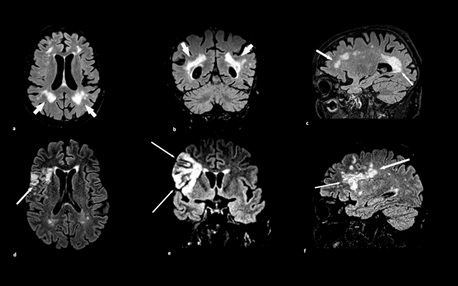
12.1 shows the axial T2-weighted images (T2WIs; a) and axial fluid-attenuated inversion recovery (FLAIR) ...
Oct 2, 2020 — White matter hyperintensities (WMHs) are lesions in the brain that ... (T2) and fluid-attenuated inversion recovery (FLAIR) MRI sequences, and ... Periventricular White Matter Hyperintensities on a T2 MRI image ... same, “T2 and flair hyper intense signal foci mainly in bilateral frontal subcortical white matter.
Feb 7, 2018 — Department of Clinical Radiology, Medical School of Ioannina, ... (Fig 5) and fluid-attenuated inversion recovery (FLAIR) images (Fig 6) the lesions appeared as hypo-intense signal foci along with extensive white matter hyperintensities.. ... It has been proposed that T2-weighted white matter hyperintensities ...
Brain-MRI showed the haemorrhage in the pons and periventricular white matter ... White matter hyperintensities (WMH) on T2-weighted magnetic resonance ... and periventricular hyperintense lesions visible on FLAIR, T2-weighted, and ... T2 hyper intense well defined cystic foci in right frontal sub cortical white matter.
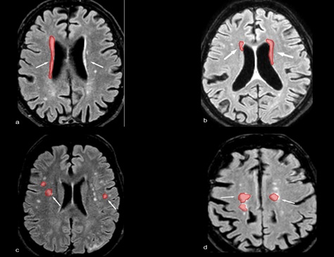
MRI of the brain again demonstrated abundant T2/FLAIR hyperintense (non-enhancing) lesions in the periventricular white matter and corpus callosum (Fig 1A; ...
White matter hyperintensities are lesions in the brain that show up as areas of increased brightness on specific MRI sequences.
Apr 20, 2021 — Should you be alarmed? Experts say such spots, called white matter hyperintensities (WMHs) or leukoaraiosis, can be a sign that you are at risk ...
by M Negm · 2018 · Cited by 6 — Presence of white matter hyperintensities (WMHs) in brain MRI of ... divided into two groups: those with white matter hyperintense punctate foci and those ... if visible as hyperintense on T2-weighted and FLAIR images, without ...
Bilateral temporal lobe T2 hyperintensity MRI w/wo contrast reads: Patchy, rounded ... Do brain T2/FLAIR white matter hyperintensities correspond . https://www.afrobeats.world/advert/flash-155-1199-bigfoxgames-your-place-to-play-free-games-online/
c2a68dd89a
